




Panoramic Dental X-Ray in Astana
In our clinic, panoramic radiography (orthopantomogram) is prescribed when indicated, specifically when it is necessary to obtain a general overview of the condition of the dentofacial system or to monitor the results of procedures performed. Diagnostics are carried out using modern equipment with a low radiation load.
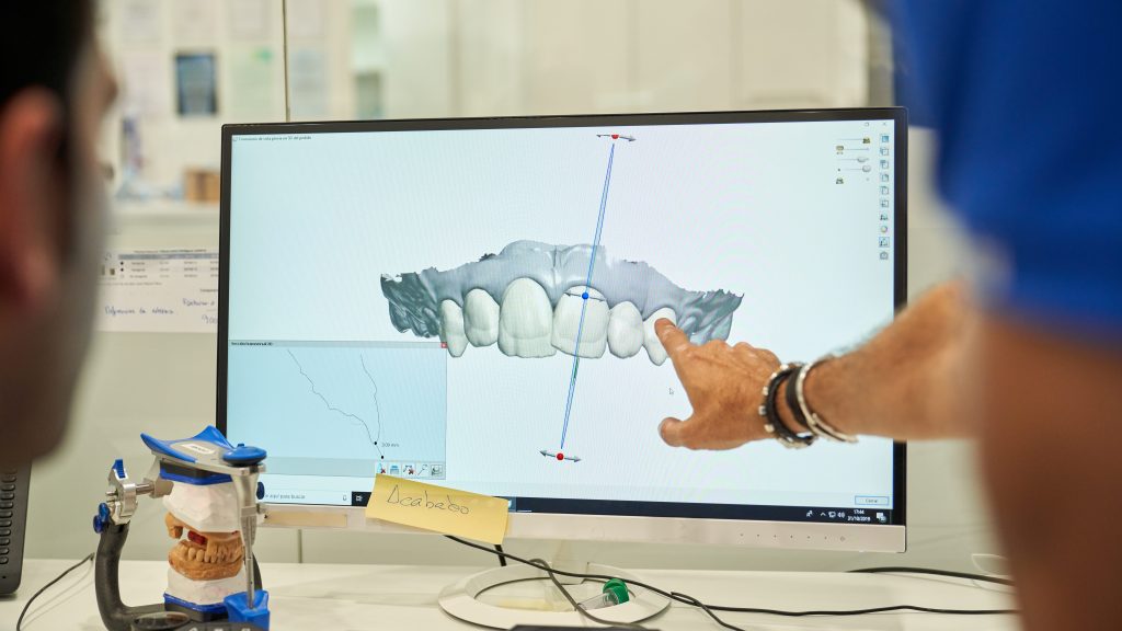
When choosing an examination method, the dentist takes into the account the specific task at hand. In some cases, a local radiograph is sufficient, while in others an orthopantomogram or three-dimensional visualization using CBCT is required. Visual images are additionally processed by the 3D Dental artificial intelligence system, which helps detect diagnostically significant features and clarify further strategy.
What an Orthopantomogram Shows
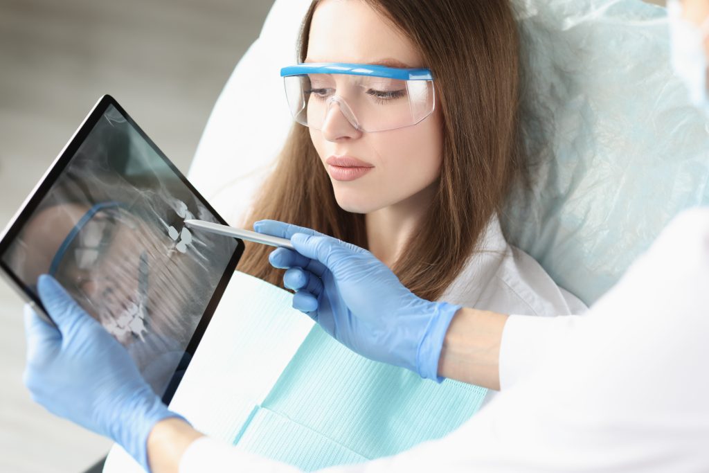
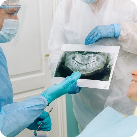
It represents a two-dimensional image that simultaneously displays the dentition, both jaws, the temporomandibular joint areas, and partially the maxillary sinuses. Thanks to the wide coverage, it is possible to evaluate the location of individual anatomical structures and see the big picture.
At Doctor Dent in Astana, this type of examination is used, for example, after implant placement, to evaluate the position of unerupted units, or for preliminary analysis before dental treatment.
At the same time, a panoramic radiograph is not intended for the detailed study of small areas. It is inferior to three-dimensional tomography in terms of the accuracy of displaying root canals, adjacent bone, and hidden inflammation. Therefore, when preparing for implantation, surgical procedures, or complex endodontics, a specialist may recommend CBCT.
It is performed on digital equipment — an orthopantomograph. Depending on the machine model and the selected mode, the radiation load remains medium or very low. The obtained data is evaluated by a specialist, and 3D Dental software analysis is used as an additional tool and does not replace a doctor’s conclusion.
When Panoramic Radiography Is Prescribed
Despite the widespread use of CBCT, this diagnostic method retains its value in many situations. The main indications can be:
- primary assessment of the dentofacial system;
- determining the position of impacted and displaced units;
- preparation for orthodontic correction;
- monitoring the movement of tooth units;
- assessment of the jaw joint area;
- detecting changes in the region of the maxillary sinuses;
- post-implantation monitoring;
- examination after jaw injuries;
- analysis of the general condition before starting comprehensive therapy.
In our clinic, it is most often used as a survey or monitoring method of visualization. For an accurate assessment of anatomy and planning complex procedures, preference may be given to cone-beam computed tomography.
Advantages of OPG at Doctor Dent
- Accurate Equipment
Technical equipment is systematically inspected and maintained in working order. Automatically configured parameters help obtain a high-quality image and reduce the likelihood of a repeat procedure. The digital format allows zooming in on specific areas, adjusting contrast, and carefully examining the area of interest. - Low Radiation Load
Such an X-ray is carried out only if indicated. During the procedure, protective equipment provided by protocol is used, including a lead apron. Equipment parameters are selected taking into account the patient’s age and the diagnostic task. - Rapid Image Acquisition
The orthopantomogram appears on the screen almost immediately after scanning. The dentist can evaluate it and adjust the restoration plan in the shortest possible time. The file is attached to the patient’s electronic medical record. This simplifies monitoring over time and allows comparing conditions at different stages.
How the Procedure Works
Special preparation is usually not required. Before scanning, the patient is asked to remove metal jewelry and items that may fall into the field of view. Then a protective apron is put on, and the patient is helped to take the correct position in the machine.
During the operation of the orthopantomograph, it is necessary to remain still. The scanning element moves around the head and forms a single image of both jaws. The entire procedure takes a few minutes and is not accompanied by painful sensations.
OPG and CBCT: What Is the Difference?
Radiography creates a two-dimensional picture. It makes it possible to see a large part of the jaw, but individual structures can overlap each other.
CBCT forms a three-dimensional model, allowing the jaw to be evaluated in different projections. Tomography makes it possible to examine in detail the position of roots, bone thickness, channel location, and other anatomical features.
Therefore, the choice depends not on which method is “better” in general, but on the task set. A panoramic view may be sufficient for a general overview, while at the stage of drawing up an implantation or surgical plan, a more detailed three-dimensional study is usually required.
Cost of Panoramic Dental X-Ray in Astana
You can find out the price of the procedure in the clinic’s price list. Depending on the service conditions, the cost may include:
- performance on a specialized machine;
- saving the image in the patient’s record;
- software processing using artificial intelligence technology;
- explanation of examination results if a consultation is included.
Prices can be specified with the receptionists.
Frequently Asked Questions
How does a panoramic image differ from a targeted intraoral X-ray?
Both methods belong to two-dimensional radiography, but differ in the area of coverage. A local radiograph shows a small area — usually one or several adjacent units. It is used for a comprehensive assessment of roots, canals, and surrounding tissues. OPG displays both jaws, dentition, and adjacent anatomical structures. It gives a less detailed, but broader picture. The appropriate option is determined by the dentist.
Can OPG be performed on children?
Radiography for young children is available in cases of urgent necessity. The specialist uses a mode with minimal radiation load.
Is the examination performed during pregnancy?
During pregnancy, any radiological examinations require a particularly well-founded prescription. In the absence of urgency, they can be postponed. In an emergency situation, the decision is made by the doctor, taking into account the gestational age, the patient’s condition, and the need for diagnosis.
Can I get the image in digital format?
Materials are stored in Doctor Dent’s digital archive and added to the patient’s medical record. If necessary, the file can be provided in a suitable format to save it or pass it to another specialist for consultation.
Booking a Panoramic Dental X-Ray in Astana
To choose a convenient time, call the number on the Doctor Dent website or request a callback. The receptionist will book your appointment and answer questions related to visiting the clinic.
The final decision on the need for an OPG is made by the dentist. This approach avoids unnecessary procedures and allows choosing the exact diagnostic method that corresponds to the clinical situation.
Blog
Expert Insights on Dental Health
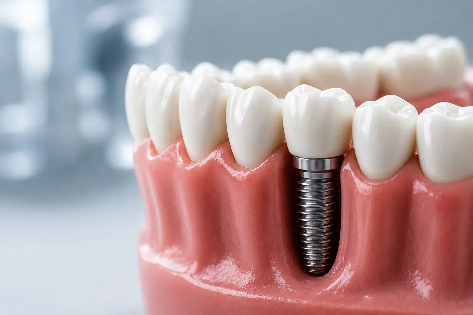
Implantation is considered one of the most reliable ways to restore lost teeth. When planning…
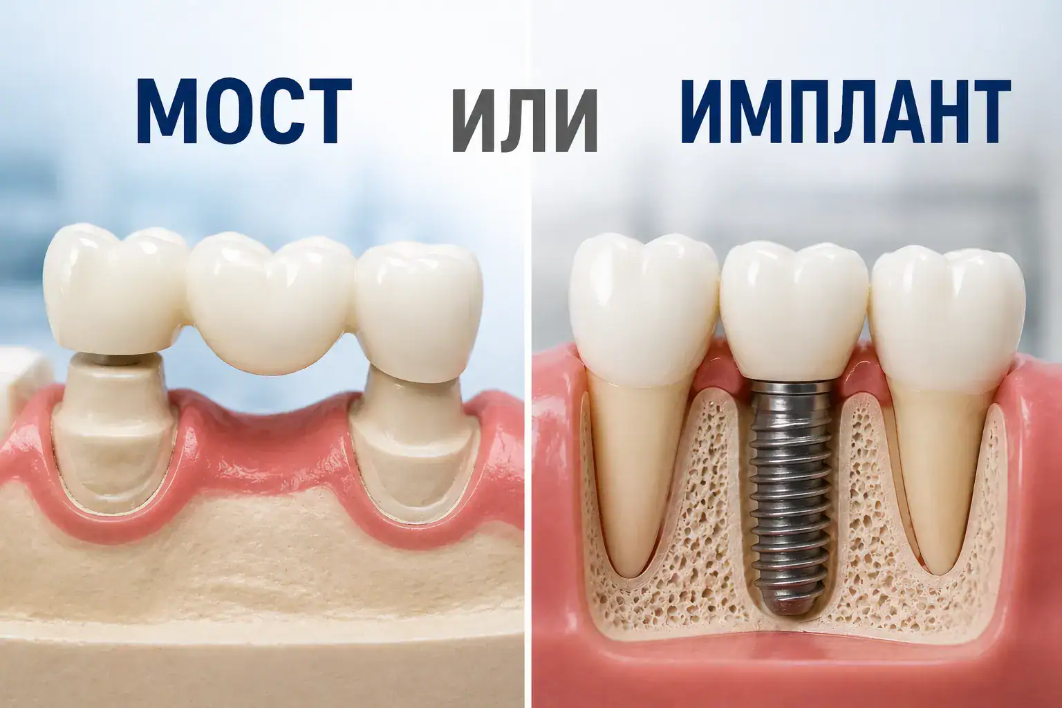
Which is better: a bridge or an implant?
Losing even a single tooth can lead not only to aesthetic discomfort but also to…

Dental Implants: Should You Be Afraid?
Dental implantation in Astana is the most sought-after procedure for restoring the integrity of the…

Caring for Braces After Installation
Braces help straighten the teeth, create a beautiful smile, and correct bite defects. These appliances…


